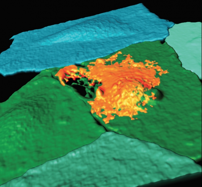
When you hear “gas-filled bubbles,” lifesaving medicine probably isn’t the first thing that comes to mind. But that’s the idea behind a recent Proceedings of the National Academy of Sciences paper from the lab of Flordeliza Villanueva, MD vice chair for preclinical research for the University of Pittsburgh Department of Medicine, director of the Center for Ultrasound Molecular Imaging and Therapeutics, and professor of medicine. Pitt postdoc Brandon Helfield was the paper’s first author.
Villanueva’s research on microbubbles has made headlines in the past. These gas-filled, smaller-than-red-blood-cell-size spheres are stimulated with ultrasound technology. To minimize side effects, the bubbles can be designed to transport therapies to specific biological sites as they move through the bloodstream. This noninvasive sonoporation, as it’s called, has been used experimentally to break up blood clots and deliver drugs to the brain. It might also be used to stem tumor growth or identify early signs of cardiovascular disease.
Now, “we’re able to get more of a granular look at what’s going on that underlies these observations people have made on a whole-organ level,” Villanueva explains.
